Learning the Origins and Insertions of Muscles may feel like a big overwhelming revision topic, however, it doesn’t have to be. As part of your Level 3 Anatomy and Physiology Exam, there are 50 muscles, each with an origin, insertion, name, location, and muscle action to learn and remember.
In this blog we share everything you need to know about the origins and insertions of muscles, and how to learn them, without overwhelm or memory blanks.
We even show you how to unpick complex exam-style questions and test your knowledge
What are Origins and Insertions?
Skeletal muscle is attached to the bone on its ends by way of what we call tendons.
As the muscles contract, they exert force on the bones, which help to support and move our body along with its appendages.
In most cases, one end of the muscle is fixed in its position [the origin], while the other end moves during contraction [the insertion].
The origin is the attachment site that doesn’t move during contraction, while the insertion is the attachment site that does move when the muscle contracts.
When we talk about origins and insertion, we also need to know some basic anatomy and physiology terms relating to bones.
Terminology for learning muscles:
Terms such as distal and proximal:
Distal being situated away from the centre of the body and proximal situated nearer to the centre of the body or the point of attachment.
The insertion is usually distal, while the origin is proximal, relative to the insertion.
For example, one could say the wrist is distal to the elbow. Conversely, you can say the elbow is proximal to the wrist.
As well as proximal and distal, you also need to know other anatomical terminology including:
Medial = The point closest to the midline of the body
Lateral = The point furthest from the midline of the body
Superior = The uppermost part
Inferior = The lowest part
Anterior = The front
Posterior = The back
Let’s dive a little deeper…
Muscular contraction produces an action, or a movement of the appendage. We will use examples to describe how the origin and insertion affect the action of a skeletal muscle.
Muscle contraction results in different types of movement.
There are several different types of movements, including flexion, extension, hyperextension, abduction, adduction, and circumduction to name a few.
[Note: you MUST be familiar with joint movements for your Level 2 and 3 Anatomy and Physiology exam]
Understanding joint movements also help you learn origins and insertions.
The movement is a direct result of muscle attachment. Most of these movements are realized when we run. Each of these actions can be described in one of two ways.
The first describes an action in terms of the bone to which the muscle is attached or the appendage that is moved.
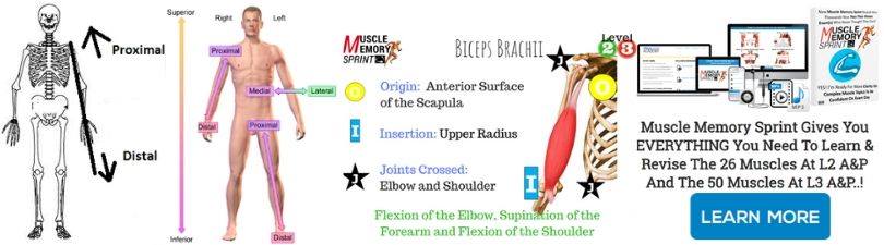
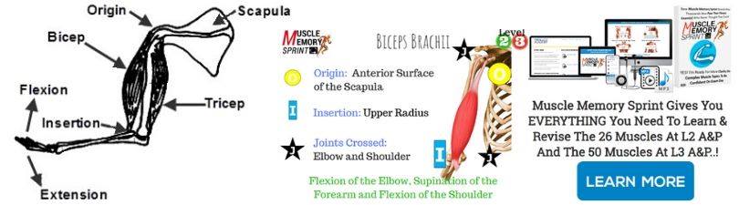
For example, the biceps brachii performs flexion of the forearm as the forearm is moved.
The second way to describe a muscle’s action is based on the joint or the articulation. For example, that same muscle, the biceps brachii, performs flexion at the elbow, in which the elbow is the joint.
Let’s stay with the biceps,
The bicep has two (bi = two) origins, one high on the humerus, the other on the scapula. When the muscle contracts and shortens it pulls on the insertion points on the radius causing the elbow to flex.
The key to learning the origins and insertions of muscles is:
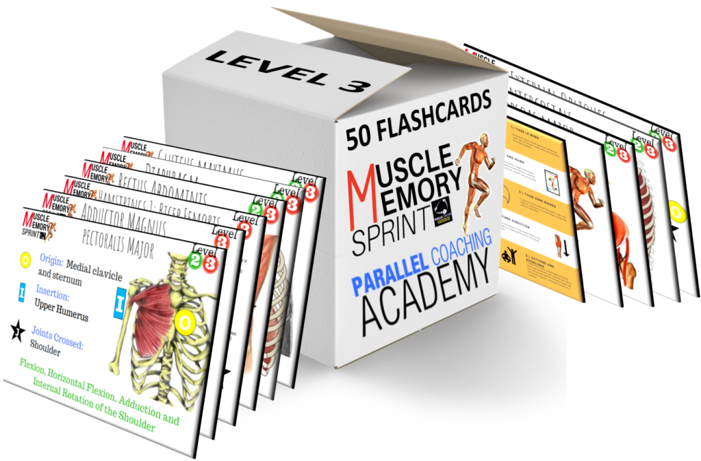
[There are 50 muscles you must know at Level 3 Anatomy and Physiology, all of which are taught in our Muscle Memory Sprint]
✔️ Knowing the muscle name and its location in the body
for example, the biceps are the upper arm and anterior of the body
✔️ Knowing the joint(s) the muscle crosses
for example, the biceps cross the Shoulder and elbow
✔️ Understanding that origins are above the joint or proximal end of the bone intended to move
for example, the biceps cross the Shoulder and originates on the scapula
✔️ Understanding that insertions are below the joint or distal end of the bone intended to move
for example, the biceps cross the elbow and inserts on the radius
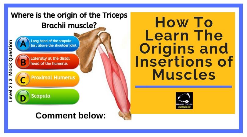
A Level 3 Anatomy and Physiology question for origins and insertions would look like:
Q: Where is the origin of the biceps brachii muscle?
- Scapula
- Lower Humerus
- Laterally at the distal end of the humerus
- Long head of the scapula just above the shoulder joint
We know from the above text that it can’t be answer B and C. These are related to lower or distal end of the humerus and the we are concerned here for the origin.
Answer D – there is no long head of the scapula! 😊
Therefore, the answer must be A – the scapula
Let’s have another go:
What muscle inserts onto the Femur?
A. Biceps brachii
B. Iliopsoas Major
C. Gastrocnemius
D. Soleus
This question is concerned with a muscle that inserts on the femur! Reading the question and reframing it is key.
What exactly is the question asking?
Staying with the questions and not looking at the possible answers…
Let’s look at what we already know and the facts,
✔️ We know the femur is the upper leg
✔️ We know the femur articulates with the pelvis to create the hip joint and with the tibia and tibia to create the knee joint.
✔️ The proximal end of the femur is closest to the hip joint and the distal end is closest to the knee joint.
✔️ We know we are looking for an insertion on the femur – therefore the origin is proximal of this and closer towards the main body/heart.
✔️ We now know we are concerned with muscles that cross the hips and/or in the upper leg – namely the quadriceps, adductors, abductors, Hip flexors and hamstring group muscles.
When we analyze the 4 possible answers,
A. Biceps brachii
B. Iliopsoas Major
C. Gastrocnemius
D. Soleus
We can immediately rule out A as we are concerned with a muscle in the upper leg, not the arm.
C and D are lower leg muscles however we can’t rule them out straight away without a quick analysis.
We know both these muscles [C and D] are concerned with the knee and ankle joint and therefore their insertions are below the ankle joint on the calcaneus – nowhere near the femur.
Therefore, the answer must be B – the Iliopsoas Major
Read the question and reframe it!
What exactly is the question after? Are you looking for an origin or insertion?
What do I know about this muscle: the name, location, joint crossed, and whether it’s the proximal or distal end of the bone?
Here are a few more origin and insertion questions to tackle – make sure to drop your answers below in the comments box:
Origins and Insertions of Muscles Mock Questions
1. Where does the Anterior Tibialis Originate?
A. Femur
B. Tibia
C. Ulna
D. Clavicle
2. What is the Insertion of the Latissimus Dorsi?
A. Base of the Skull
B. The Thoracolumbar Aponeurosis
C. The Upper Humerus
D. The Iliac Crest
3. What muscle originates on the Inferior edge of the Skull
A. Trapezius
B. Pectoralis Major
C. Biceps Brachii
D. Biceps Femoris
Download More Mock Questions for FREE just link these by clicking this link: MOCK QUESTIONS
The answers are:
Q1 = A, Q2 = C, Q3 = A
How to learn orgins and insertions?
Learning all of the muscles can feel overwhelming and frustrating. Staring at a manual and learning from the smallest of images.
Here’s How Any Trainee Fitness Professional Can Remember Everything To Do With Muscles In As Little As 15-Minutes Every Day (Without Getting Overwhelmed Or Relying On Your Course Manual)
- Gives You The Confidence To Do Well On Any Muscle Question
- How To Learn EVERY Muscle You Need To Know For Your Exam
- Escape From The Worry And Stress About Muscle Questions And Failing Your A&P Exam
- Makes It Easy To Crush Your A&P Exam
https://revision.parallelcoaching.co.uk/level-3-muscle-memory-flashcards
Dedicated to More
Hayley “Origins and Insertions of Muscles” Bergman
Parallel Coaching
P.S. You can also find us on the following platforms:
Instagram: Follow Now
Facebook: Like Our Page
Twitter: Tweet Us
YouTube: Subscribe Here
More Anatomy Revision Blogs: HERE