If you are studying for your Level 2 or Level 3 Anatomy and Physiology exam, then you need to know the structure of a long bone
This blog will teach you about the structure of the long bone, and you can check your knowledge with a quiz at the end. Before we dive into the detail of a long bone, let’s zoom out a little and understand the basics of the skeletal system:
Key Skeletal System Facts:
- The typical human skeleton consists of 206 bones in adults.
- More bones are present at birth, gradually fusing together as the body matures.
- The skeleton is divided into two parts…
You need to know about Axial and Appendicular Skeletons for your exam…
The axial skeleton and the appendicular skeleton
The axial skeleton includes the bones of the skull, face, and spine along with the ribs and breastbone.
The appendicular skeleton includes the bones of the arms, hands, legs, feet, and pelvis as well as the clavicles and shoulder blades.
An easy way to remember the axial and appendicular skeleton is the ‘Law of the ones and twos’ – If the body only has one of the bones such as the skull then it’s part of the axial skeleton. Whereas if the body has two of the same bones then it’s part of the appendicular skeleton such as the tibia.

You won’t find any long bones in the axial skeleton!
Long bones — a subtype of bones — are longer than they are wide.
These are strong bones because they must be able to withstand the force generated when the body moves and changes direction.
Though different long bones have different shapes and functions, they all have the same general structure. Examples of long bones include the femur, tibia, radius and ulna.
Let’s breakdown the structure of a long bone
Articular cartilage is the smooth, white tissue that covers the ends of bones where they come together to form joints. Healthy cartilage in our joints makes it easier to move. It allows the bones to glide over each other with very little friction. Articular cartilage can be damaged by injury or normal wear and tear.
Every long bone is capped with wide areas on each end which are called epiphyses. The epiphysis closer to the torso is called the proximal epiphysis while the distal epiphysis is at the farther end. Epiphyses are filled with spongy bone containing red bone marrow, which is red in color because it makes red blood cells. Each epiphysis is capped with articular cartilage that connects the bone to the rest of the body while simultaneously cushioning the end of the bone.
The largest part of any long bone is the long cylindrical middle, called the diaphysis. The diaphysis takes the brunt of the force a long bone must support and is made up primarily of compact bone — a dense, strong bone composed of minerals, including calcium, phosphorus, and magnesium, as hard as many types of rock. The diaphysis also has small holes for blood vessels that carry nutrients to the compact bone cells.
What types of bone are in a long bone?
Spongy bone, also known as cancellous bone or trabecular bone, is a very porous type of bone. Spongy bone is usually located at the ends of the long bones (the epiphyses), with the harder compact bone surrounding it.
Compact bone, also known as cortical bone, is a denser material used to create much of the hard structure of the skeleton. As seen in the image below, compact bone forms the cortex or hard outer shell of most bones in the body. The remainder of the bone is formed by cancellous or spongy bone.
The periosteum is a membrane that covers the outer surface of all bones, except at the joints of long bones.
Endosteum lines the inner surface of the medullary cavity of all long bones.
An epiphyseal disk of cartilage at the junction of the diaphysis and epiphyses of growing long bones. Cartilage synthesis provides for growth in length; eventually, the cartilage is replaced by bone.
The medullary cavity (medulla, innermost part) is the central cavity of bone shafts where red bone marrow and/or yellow bone marrow (adipose tissue) is stored; hence, the medullary cavity is also known as the marrow cavity.
There are two types of bone marrow: red marrow (also known as myeloid tissue) and yellow marrow. Red blood cells, platelets, and most white blood cells arise in red marrow; some white blood cells develop in yellow marrow. The colour of yellow marrow is due to the much higher number of fat cells.
Red marrow is present in adulthood only in the flat bones of the skull, the sternum, ribs, vertebral column, clavicle, humerus, and part of the femur. Notice not all long bones have red marrow.
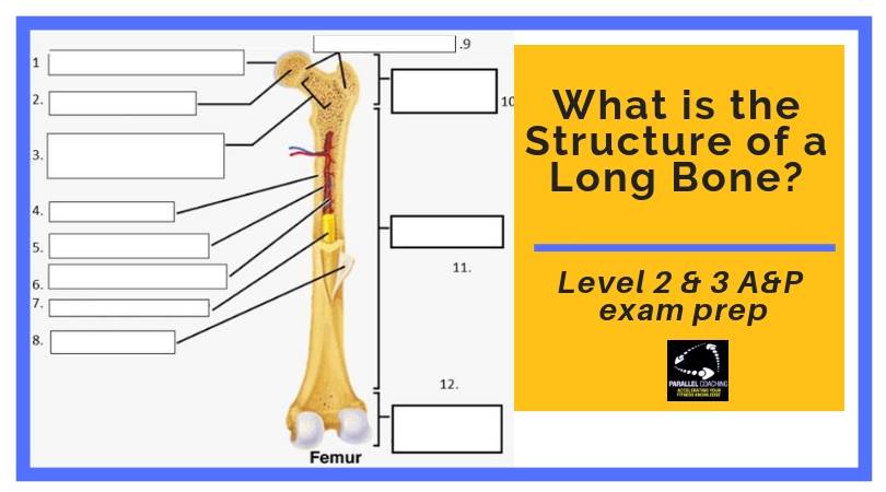
Now you are familiar with the 12 features of a long bone – Test yourself below and fill in the blanks.
[Note the answers are at the bottom of this blog!]
Test Your Knowledge: Structure of a Long Bone Quiz:
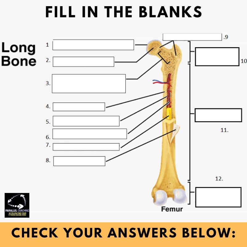
Mock Question Answers:
1. Articular Cartilage
2. Spongy Bone
3. Space occupied by red bone marrow
4. Endosteum
5. Compact Bone
6. Medullary Bone
7. Yellow Marrow
8. Periosteum
9. Epiphyseal Discs
10. Proximal Epiphysis
11. Diaphysis
12. Distal Epiphysis
If you want more mock questions like this, then you can download more Free Mock Questions: DOWNLOAD NOW
Need More Help with your Level 2 Anatomy Revision?
For Trainee FITPROS Taking Their L2 Anatomy & Physiology Exam.
Learn, Revise & Pass Your Level 2 Anatomy & Physiology Exam In Under 10-hours
(Without Having To Spend Hours Revising Or Feeling Overwhelmed)
If you want to get your revision structured, learn everything you need to know, and feel confident on exam day, then click the link below:

Dedicated to More
Hayley “structure of a long bone” Bergman
Parallel Coaching
P.S. You can also find us on the following platforms:
Instagram: Follow Now
Facebook: Like Our Page
Twitter: Tweet Us
YouTube: Subscribe Here
More Skeletal System Blogs: HERE