
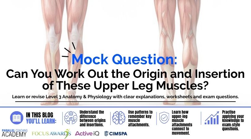
Struggling to remember muscle origins and insertions? Learn how to understand upper-leg muscle attachments, actions and exam questions.
You know the muscle.
You know what it does.
But then the exam asks:
“What are the origins and insertions of this muscle?”
And suddenly, everything you’ve learned seems to disappear.
Was it the femur?
The tibia?
The pelvis?
And which end was the origin again?
If this sounds familiar, you’re not alone.
Origins and insertions can feel like a huge list of random anatomical facts that you’re simply expected to memorise.
But there’s a better way to approach them.
Don’t just memorise the attachment. Understand where the muscle is, what joints it crosses and how its attachments help explain what it does.
Let’s see how.
Mock Question: Can You Work It Out?
Let’s start with a question.
Which of the following correctly identifies the origin and insertion of the rectus femoris?
A. Origin: ischial tuberosity; insertion: head of fibula
B. Origin: anterior inferior iliac spine; insertion: tibial tuberosity via the quadriceps mechanism
C. Origin: linea aspera; insertion: medial tibial condyle
D. Origin: pubis; insertion: medial surface of the tibia
Don’t worry if you’re not immediately sure.
We’ll come back to the answer.
More importantly, we’re going to look at how you could work it out.
Why Do You Need to Know Origins and Insertions?
At Level 3, you’re not just learning the names of muscles.
You’re learning how the musculoskeletal system works.
Understanding where a muscle attaches helps you understand:
- where the muscle is located
- which joints it crosses
- what movement it can produce
- why it performs certain actions
- how muscles contribute to exercise
- why particular exercises involve particular movements
CIMSPA’s Personal Trainer standard includes knowledge of muscle attachment sites, including origins and insertions, alongside muscle structure and function.
So this isn’t simply information you need to remember for an exam.
It’s part of understanding why the body moves the way it does.
And that’s where origins and insertions become much more interesting.
Origins and Insertions: What’s the Difference?
Let’s keep this simple.
Origin
The origin is generally described as the more proximal or relatively fixed attachment of a muscle.
Insertion
The insertion is generally described as the more distal or relatively movable attachment.
But don’t fall into the trap of thinking:
“The origin never moves, and the insertion always moves.”
That’s an oversimplification.
Depending on the movement, either attachment can move.
Instead, think of origin and insertion as descriptions of where the muscle attaches.
You should also understand the terms:
Proximal = closer to the trunk or point of attachment.
Distal = further away from the trunk or point of attachment.
This becomes particularly useful when you’re looking at the muscles of the upper leg.
The Simple Way to Learn Origins and Insertions
Instead of trying to memorise:
Muscle → origin → insertion
as three completely separate facts, use this sequence:
Muscle – Where is it?
↓
Joint – Which joint or joints does it cross?
↓
Origin – Where is its proximal attachment?
↓
Insertion – Where is its distal attachment?
↓
Action – What movement would those attachments allow it to produce?
This gives you a way to connect the information.
And once the information is connected, it’s much easier to remember.
Let’s try it with the muscles of the upper leg.
Start With the Quadriceps
The quadriceps femoris is made up of four muscles:
- Rectus femoris
- Vastus lateralis
- Vastus medialis
- Vastus intermedius
Together, they are the major muscles responsible for knee extension. All four contribute to the quadriceps tendon, which continues through the patella and patellar ligament to the tibial tuberosity.
So instead of learning four completely unrelated muscles, start by spotting the pattern.
The three vasti muscles are:
1.Vastus lateralis
Origin: greater trochanter and lateral lip of the linea aspera of the femur.
Insertion: quadriceps tendon → patella → patellar ligament → tibial tuberosity.
2. Vastus medialis
Origin: intertrochanteric line and medial lip of the linea aspera of the femur.
Insertion: quadriceps tendon → patella → patellar ligament → tibial tuberosity.
3. Vastus intermedius
Origin: anterior and lateral surfaces of the femoral shaft.
Insertion: quadriceps tendon → patella → patellar ligament → tibial tuberosity.
Notice the pattern?
All three vasti- muscles:
- Originate from the femur.
- Contribute to the quadriceps mechanism.
- Cross the knee.
- Extend the knee.
That’s much easier to remember than three separate lists.
Then There’s Rectus Femoris – origins and insertions
Rectus femoris is the interesting one.
Why?
Because unlike the three vasti, it crosses both the hip and knee joints.
Its origin is on the pelvis, including the anterior inferior iliac spine, and it contributes to the same quadriceps mechanism at the knee.
That gives you another useful connection:
Origin on pelvis
↓
Crosses hip
↓
Crosses knee
↓
Inserts through quadriceps mechanism
↓
Hip flexion + knee extension
Suddenly, the actions make more sense.
It’s not just:
“Rectus femoris does hip flexion and knee extension because my textbook told me.”
You can understand why.
Pattern Spotting Is Your Friend
Here’s an important revision technique.
When you’re learning a group of muscles, don’t immediately ask:
“What are all the individual origins and insertions?”
First ask:
What do they have in common?
For the quadriceps:
- Three vasti originate from the femur.
- Rectus femoris originates from the pelvis.
- All four contribute to knee extension.
- Rectus femoris also crosses the hip.
Those patterns give you something to hang the detailed information onto.
Now Let’s Look at the Hamstring Origins and Insertions
The main hamstring muscles are:
- Biceps femoris
- Semitendinosus
- Semimembranosus
Biceps femoris has a long head and a short head.
Here’s another great example of why understanding anatomy is better than simply memorising a table.
The long head of biceps femoris, semitendinosus and semimembranosus have proximal attachments around the ischial tuberosity, while the short head of biceps femoris originates from the femur.
That’s important.
Because the short head is different.
Biceps femoris
Long head
Origin: ischial tuberosity.
Insertion: head of the fibula.
Short head
Origin: lateral lip of the linea aspera and lateral supracondylar region of the femur.
Insertion: head of the fibula.
Semitendinosus
Origin: ischial tuberosity.
Insertion: medial proximal tibia.
Semimembranosus
Origin: ischial tuberosity.
Insertion: medial condyle of the tibia.
Again, look for the pattern.
The main hamstrings, apart from the short head of biceps femoris, cross both the hip and knee.
That helps explain their major actions:
Hip extension
and
Knee flexion.
Here’s the Pattern You Should Notice
Compare the quadriceps and hamstrings.
Quadriceps – Mostly on the front of the thigh. Main action – Knee extension
Hamstrings – On the back of the thigh. Main actions – Hip extension + knee flexion
This isn’t just useful for remembering actions.
It helps you understand the attachments.
If a muscle crosses the knee and is positioned anteriorly, you can start thinking about how its line of pull relates to knee extension.
If it crosses posteriorly, you can start thinking about knee flexion.
Location + joint crossed + attachment + action = connected knowledge.
What about origins and insertions of Other Upper Leg Muscles?
You don’t need to learn every muscle of the thigh as one enormous list.
But there are some useful examples that help you build the bigger picture.
Sartorius
Origin: anterior superior iliac spine.
Insertion: medial surface of the proximal tibia.
Because it crosses the hip and knee, it can contribute to hip flexion and knee flexion, as well as movements involving rotation of the thigh and leg.
Gracilis
Origin: pubis.
Insertion: medial proximal tibia.
It crosses both the hip and knee and contributes to hip adduction and knee flexion.
Adductor longus
Origin: pubis.
Insertion: linea aspera of the femur.
Its position helps explain its primary role as a hip adductor.
These examples also appear within the broader group of muscle attachments included in CIMSPA’s Personal Trainer standard.
Again, don’t simply memorise the facts.
Look at where the muscle starts, where it finishes and which joints it crosses.
What If You Can’t Remember the Answer in an Exam?
This is where understanding becomes really useful.
Imagine you’re given a question about a muscle and you can’t immediately remember its origin.
Don’t panic.
Try working backwards.
Step 1: Where is the muscle?
Is it on the front, back or inside of the thigh?
Step 2: What joint does it cross?
Hip?
Knee?
Both?
Step 3: Where could its attachment be?
Pelvis?
Femur?
Tibia?
Fibula?
Step 4: Does the proposed action make sense?
If the answer says a muscle performs an action that doesn’t fit its position or the joints it crosses, you’ve potentially found the wrong answer.
You’re not magically recreating the entire anatomy textbook from memory.
You’re using what you know to narrow down the possibilities.
That’s a much stronger exam skill.
Origins + Insertions + Action = Connect the Dots
Let’s take rectus femoris again.
You could memorise:
Origin: AIIS
Insertion: tibial tuberosity via the quadriceps mechanism
Action: hip flexion and knee extension
Or you could understand the relationship.
It originates from the pelvis.
↓
It crosses the hip.
↓
It continues down the front of the thigh.
↓
It crosses the knee.
↓
It attaches through the quadriceps mechanism.
↓
It can therefore contribute to movement at both joints.
That gives you a mental picture.
And mental pictures are often easier to recall than isolated words.
Why Does This Matter as a Personal Trainer?
This knowledge shouldn’t stop at the exam.
Imagine you’re teaching a squat.
You don’t necessarily need to give your client a lecture about the origins and insertions of the quadriceps.
But you need to understand what is happening.
When you’re selecting exercises, analysing movement or explaining why an exercise involves particular muscles, your knowledge of anatomy helps you make better decisions.
For example, understanding that the hamstrings cross both the hip and knee helps you understand why their role isn’t simply:
“They bend the knee.”
They also contribute to hip extension.
That becomes relevant when you’re considering exercises and movements involving both joints.
A&P isn’t supposed to live in your workbook.
It’s there to help you understand what you’re seeing in front of you.
Common Exam Mistake: Learning the List Without Learning the Location
One of the biggest problems with origins and insertions is trying to learn them as flashcards with no anatomical picture behind them.
You might remember:
“Biceps femoris — ischial tuberosity — fibular head.”
But then the exam phrases the question differently.
Suddenly, you’re stuck.
Instead, try to picture the muscle.
- Posterior thigh.
- Starts around the pelvis for the long head.
- Travels down the lateral side of the thigh.
- Crosses the knee.
- Attaches at the fibular head.
Now you’ve got a story.
And stories are easier to remember than disconnected facts.
Don’t Fall Into the Multiple-Head Trap
Another common source of confusion is assuming that every muscle has one simple origin and one simple insertion.
Some muscles have different heads or multiple attachment sites.
Biceps femoris is a great example.
The long head and short head have different origins, but they come together distally to insert around the head of the fibula.
That means when you see a question asking about biceps femoris, you need to pay attention to which head the question is referring to.
This is exactly why understanding the anatomy is more useful than memorising a single simplified sentence.
The Muscle Memory Framework to Use When Revising
If you’re revising origins and insertions, use this sequence every time:
1. Find the muscle
Where is it?
2. Find the joints
Which joint or joints does it cross?
3. Find the origin
Where is the proximal attachment?
4. Find the insertion
Where is the distal attachment?
5. Find the action
What movement does the line of pull help create?
6. Find the pattern
What does this muscle have in common with others?
This gives you six connections instead of one fact to memorise.
And that’s the difference between knowing a list and understanding anatomy.
Test Yourself: 5 More Mock Questions
Ready?
Try answering these without looking back through the article.
Question 1
Which muscle originates from the anterior inferior iliac spine and contributes to knee extension?
A. Rectus femoris
B. Semitendinosus
C. Biceps femoris
D. Gracilis
Question 2
Which muscle has an origin on the linea aspera and inserts onto the head of the fibula?
A. Semimembranosus
B. Vastus medialis
C. Biceps femoris short head
D. Sartorius
Question 3
Which hamstring muscle inserts onto the medial proximal tibia?
A. Biceps femoris
B. Semitendinosus
C. Vastus lateralis
D. Rectus femoris
Question 4
Which muscle originates from the pubis and inserts onto the medial proximal tibia?
A. Sartorius
B. Gracilis
C. Vastus intermedius
D. Biceps femoris
Question 5
Which statement about the hamstrings is correct?
A. All hamstrings originate from the femur.
B. All hamstrings cross both the hip and knee.
C. Most hamstrings cross both the hip and knee, but the short head of biceps femoris is an exception.
D. The hamstrings are primarily responsible for knee extension.
Answers
1. A
2. C
3. B
4. B
5. C
Want More Level 3 Anatomy & Physiology Practice?
Origins and insertions can feel like one of those topics where you either remember the answer or you don’t.
But you don’t have to approach them that way.
The more you understand where a muscle is, which joints it crosses, where it attaches and how that relates to its action, the easier the individual facts become to remember.
And that’s exactly how we approach Level 3 Anatomy & Physiology revision at Parallel Coaching.
If you’re preparing for your Level 3 assessments, our Anatomy & Physiology Revision Bootcamp is designed to help you work through the topics that can be difficult to understand, practise exam-style questions and build your confidence before assessment.
https://courses.parallelcoaching.co.uk/products/level-3-anatomy–physiology-revision-bootcamp
Because the aim isn’t simply to memorise more information.
It’s to understand what you’re learning well enough to use it.
And when you can do that, the exam questions become much easier to tackle.
Dedicated to More
Hayley “Muscle Origins and Insertions: Upper Leg A&P Explained” Bergman
Parallel Coaching
P.S. You can also find us on the following platforms:
Instagram: instagram.com/parallelcoaching
Facebook: https://www.facebook.com/ParallelCoaching
YouTube: https://bit.ly/2F1Z1bs
Read more blogs about becoming a PT: HERE
TrustPilot Reviews: HERE